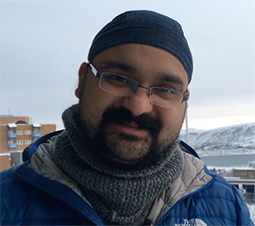
X-SIG Series: Balpreet Singh Ahluwalia
By Cameron D’Amica, Staff Writer
Dr. Balpreet Singh Ahluwalia, Associate Professor in the Department of Physics and Technology at Uit, The Arctic University of Norway, delivered two lectures at Gettysburg College for the X-SIG Seminar Series. Dr. Ahluwalia presented his public lecture Photonic Research: Its Impact to Human Society on Monday, March 4, and he presented his technical lecture, Photonic-Chip Based Optical Nanoscopy Enables Imaging of Nanoscale Biological Systems in Mara Auditorium on Tuesday, March 5.
Dr. Ahluwalia explained his field of research by outlining the research activities of the nanoscopy group at UiT. His technological development research includes optical nanoscopy, quantitative phase microscopy, optical trapping, and integrated photonics. In bioimaging, his research covers vascular biology, molecular cancer, and nanoscopy in pathology.
In order to describe his research on photonic chip-based multi-modality optical nanoscopy, he presented a background of optical nanoscopy that began with the invention of the microscope in approximately 1600, to the discovery of the diffraction limit in 1866, and the development of nanoscopy in 2005. Light diffraction and resolution, which are oftentimes confused, limited microscopy.
Dr. Ahluwalia explains how nanoscopy, super-resolution fluorescence microscopy, evolved and how people became interested in the field in the last twenty-five years. He outlined the difference between electron microscopy, which is high resolution still images, and nanoscopy, which is high resolution with real-time movie.
He continued to introduce his research on nanscopy and his photonic chip-based multi-modality optical nanoscopy, which utilizes a simple microscope and complex chip that holds the sample instead of a complex microscope with a simple glass slide to hold the sample. He describes this as the “coffee model” where a relatively cheap machine is used with an expensive product that you must “refill.”
In his chip-based optical nanoscopy, Ahluwalia explains how a waveguide chip is used as a cover slip to grow cells, and the sample is placed directly on top of waveguide. An evanescent field of optical waveguide illuminates the cell, while light is guided inside the waveguide using total internal reflection, and the intensity in the surface decays exponentially. This generates an evanescent field independent of an objective lens. This integrated platform enables easy multicolor imaging with bioimaging applications.
Chip-based nanoscopy can create 100 times larger field of view than the alternative, which is necessary for pathology where doctors need a larger sample to view. This also creates quicker results, as the chip enables fast correlative light electron imaging. There are so many biological applications of the chip-based nanoscopy that Ahluwalia is researching and developing.
At the completion of his lecture, Ahluwalia acknowledged the optical nanoscopy group at UiT that he works with and his collaborators before opening up the floor for questions on nanoscopy and his research in the field.

